▲
Fossil Preparation 5
PREPARATION OF A DINOSAUR CERVICAL VERTEBRA FROM THE ERIC THE RED WEST SITE 2016 FIELD SEASON
By Lesley Kool
Bone #287, broken cleanly into two pieces, was labelled "Probable vert from hole" and was found during the 2016 Eric the Red West field trip (image #1). Its triangular cross section and spongy internal texture suggested that it was indeed a vertebra, but from what animal?
The two pieces were in fairly large chunks of rock, so they were trimmed to make them more manageable and to cut down the preparation time. After both broken bone surfaces were consolidated with Paraloid the more robust half was prepared. The surrounding matrix was carefully chipped away from the bone using a pneumatic ARO air scribe. Most of the rock came away cleanly from the surface of the bone, but there were some rough or "rugose" areas at the edges of the bone where the individual sand grains in the rock were embedded in the bone structure. They had to be removed using a tungsten carbide needle under a low powered microscope. Newly exposed bone was treated with Paraloid to prevent it from crumbling from the vibrations caused by the ARO air scribe.
Once the first part of the bone was removed from the matrix it was glued onto the second half and the whole bone was prepared revealing the centrum (cotton-reel shaped lower part) of a dinosaur vertebra (image #3), measuring approximately 30mm in length. The internal structure of the centrum was made up of compact spongy bone suggesting that it came from an ornithopod dinosaur whereas the centra of theropod dinosaurs are much less spongy and are made up of large air-filled cavities supported by a bony lattice.
Comparison of the centrum was made with vertebrae in Peter Galton's 1974 monograph on Hypsilophodon foxii. An illustration of the cervical vertebrae (fig.20A) of H.foxii shows a marked similarity between the ventral surface of the centrum and the 3rd or 4th cervical vertebra of R196 (Stephen Poropat pers.comm). The pinched keel visible in image #4 compares well with that illustrated in figure 20A of Galton (image #5), as well as the difference in the width of the centrum, being much wider anteriorly where the cervical ribs were attached.
The dorsal view of the centrum, where the dorsal processes would have been attached, shows a series of faint parallel grooves. This indicates that possibly the dorsal processes were not fully ossified and that they were still partly cartilaginous, suggesting that the individual was a juvenile or sub-adult (teenager).
A number of vertebrae have been found at the Eric the Red West site since its discovery in 2005 and most of them have turned out to belong to small ornithopod dinosaurs. Hopefully their eventual preparation will lead to further identification of these small herbivorous dinosaurs.
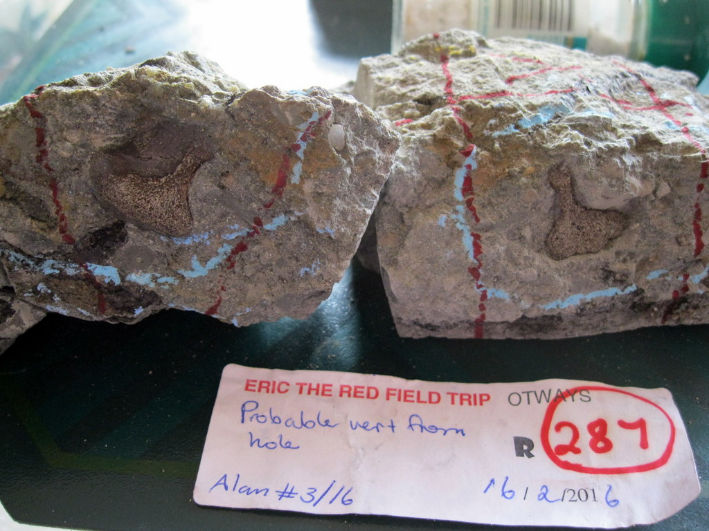
Image #1 Bone #287 shown in cross-section.
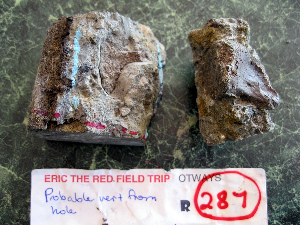
Image #2 Half the bone exposed.
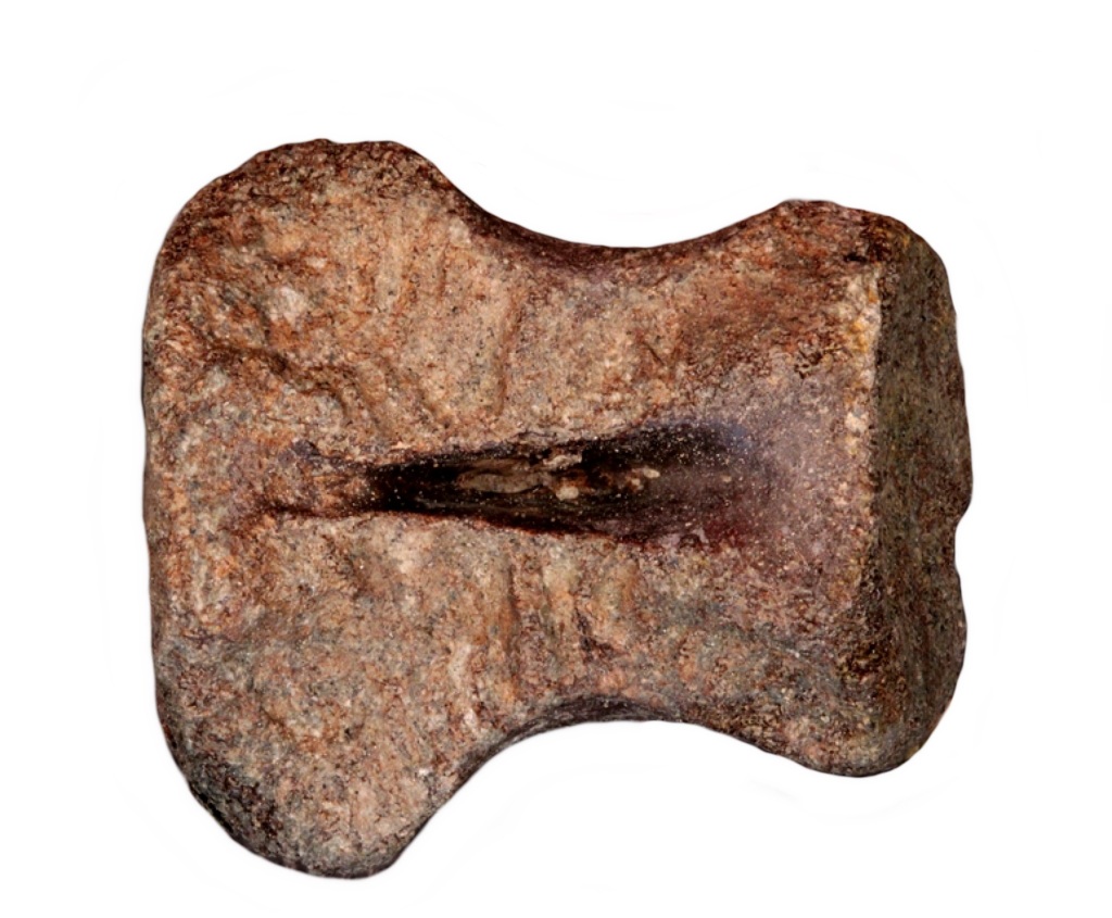
Image #3 Dorsal view of the cervical vertebra.
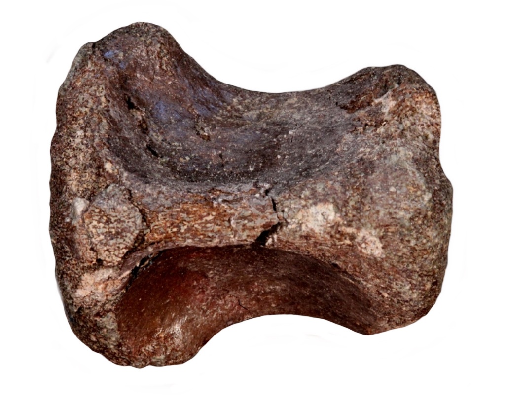
Image #4 Ventral view of the cervical vertebra showing pinched keel.
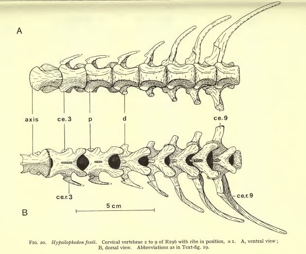
Image #5 Fig 20 from Galton's monograph on Hypsilophodon foxii.
visits