▲
Fossil Preparation 3
PREPARATION OF A SECOND DINOSAUR TIBIA FOUND AT ERIC THE RED WEST 2016
By Lesley Kool
The first dinosaur tibia, whose preparation was described last month, was partially exposed along the length of the shaft, making it readily identifiable as a small dinosaur limb. The second tibia was exposed differently in that only a cross-section through the shaft was visible (image #1). The shape of the cross-section indicated that it was a limb of some kind and that it was infilled with sediment, suggesting that one or both ends were not preserved. This is the general rule, however, occasionally a whole bone is crushed, causing the side of the shaft to cave in allowing sediment to enter the hollow cavity.
In the case of the second tibia, the first scenario turned out to be correct. Close examination of the cross-sections revealed the direction in which the bone was lying in the rock. After the rock was trimmed (image #2) with a diamond tipped rock saw, each half of the bone was exposed using the pneumatic ARO tool. One half quickly terminated in a broken end, indicating that the distal end (furthest from the body � see explanation of terminology in last month�s feature) of the bone was not preserved. Fortunately, the proximal end (closest to the body, articulating with the distal end of the femur) was well preserved and allowed us to identify the bone as a small ornithopod dinosaur tibia.
The incomplete part of the tibia was then removed from the rock and glued back onto the proximal end (image #3). Finally, the complete tibia was removed from the rock and consolidated using a weak solution of Paraloid and acetone (image #4).
The difference between the two dinosaur tibiae is quite marked. Apart from the fact that the first tibia is complete, the second tibia obviously came from a larger individual. The first tibia measures approximately 12cm in length, whereas the second tibia, missing at least half its length measures 8cm. The most interesting difference between the two bones is the shape of their proximal ends. The proximal end of the first tibia is almost rectangular in cross-section (image #5), whereas the shape of the proximal end of the second tibia is more elongate (image #6). Our initial thought was that we were looking at two different species of small ornithopod dinosaurs but in consultation with Dr Paul Barret from the British Museum of Natural History, who is an expert on ornithopod dinosaurs, he explained that there is a degree of variation in dinosaur bone articulations, often based on size. His paper on Jeholosaurus shanyuanensis*, a small ornithopod dinosaur from China, clearly indicates the difference between the tibiae of two specimens of Jeholosaurus, which mirror the shapes of the proximal ends of the two tibiae from the Eric the Red West site (images #7 and #8).
The partial dinosaur skeleton that was found at the Eric the Red West site in 2005 unfortunately does not preserve the proximal end of the tibia, however, one of the partial ornithopod dinosaur skeletons found at Dinosaur Cove during the 1980s does have the tibiae preserved (image #9) and they look remarkably like the second tibia with a wide, but narrow proximal end.
Interestingly the most common tibiae that we have found along the Bass Coast and at the Flat Rocks site, which we excavated for 20 years, are very similar to the second tibia from the Eric the Red West site. They are also similar in size, which could be the reason they have a similar morphology (image #10). We may be seeing the difference between a juvenile and an adult ornithopod dinosaur or we may have two different species of ornithopod dinosaur. Only time and more specimens will help to solve the mystery.
Next month we look at the preparation of a dinosaur vertebra.
*Feng-Lu Han, Paul M. Barrett, Richard J. Butler and Xing Xu. 2012 Postcranial anatomy of Jeholosaurus shangyuanensis (Dinosauria: Ornithischia) from the Lower Cretaceous Yixian Formation of China. Journal if Vertebrate Paleontology Vol.32:6, pp. 1370 - 1395.
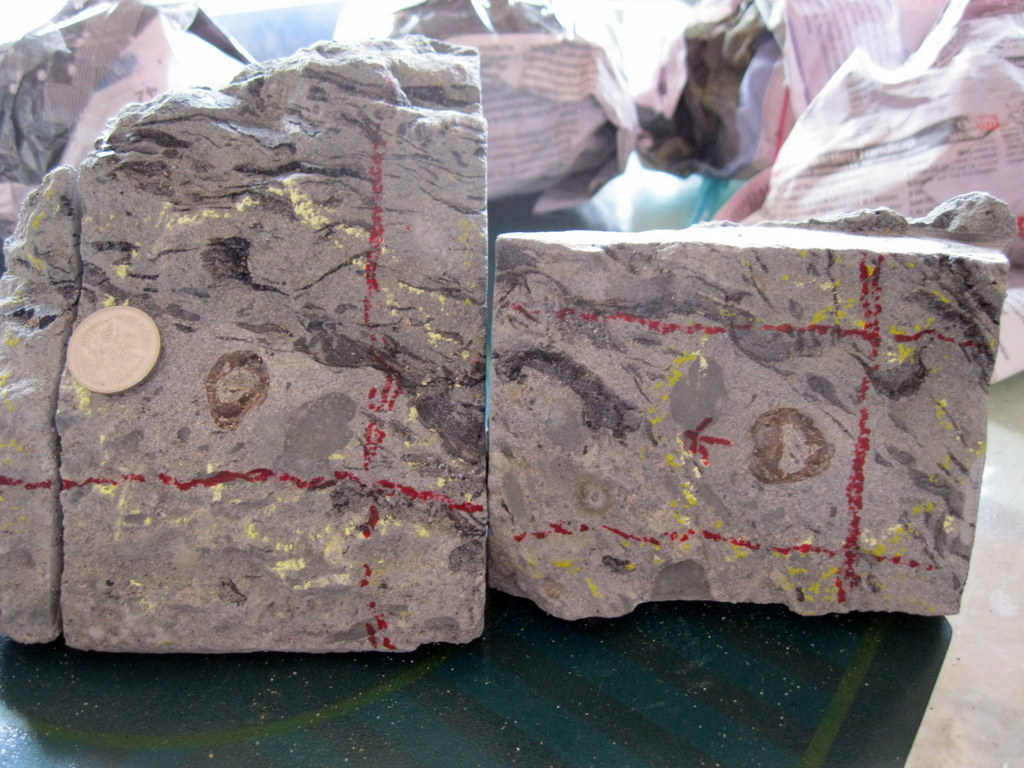
Image #1 cross-section through bone.
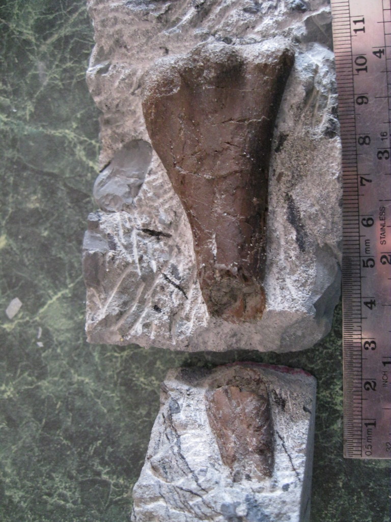
Image #2 both ends exposed.
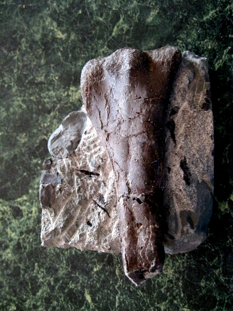
Image #3 - two halves back together.
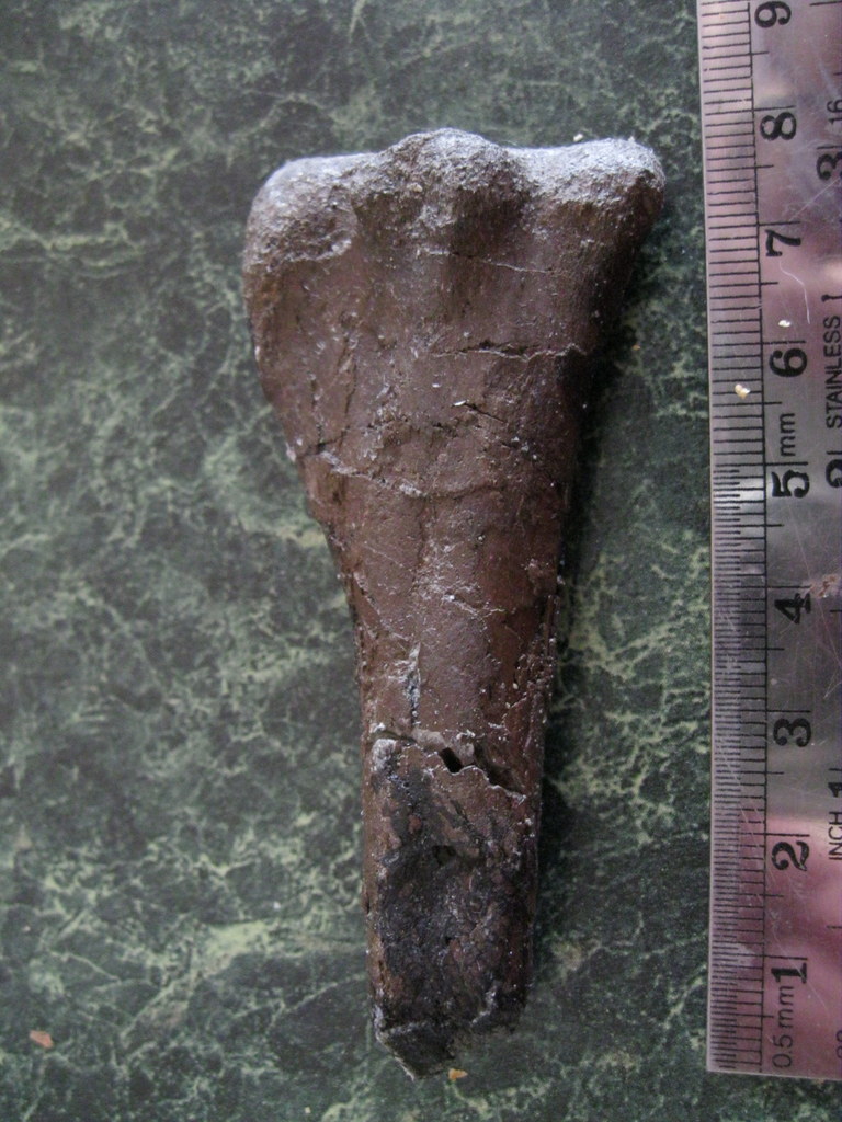
Image #4 tibia out of the rock.
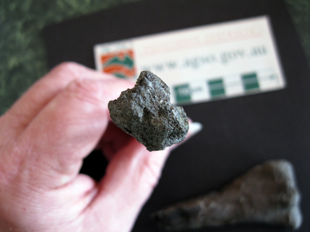
Image #5 - prox.end of 1st tibia.
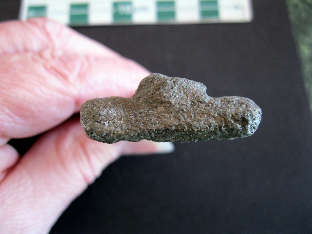
Image #6 - prox.end of 2nd tibia.
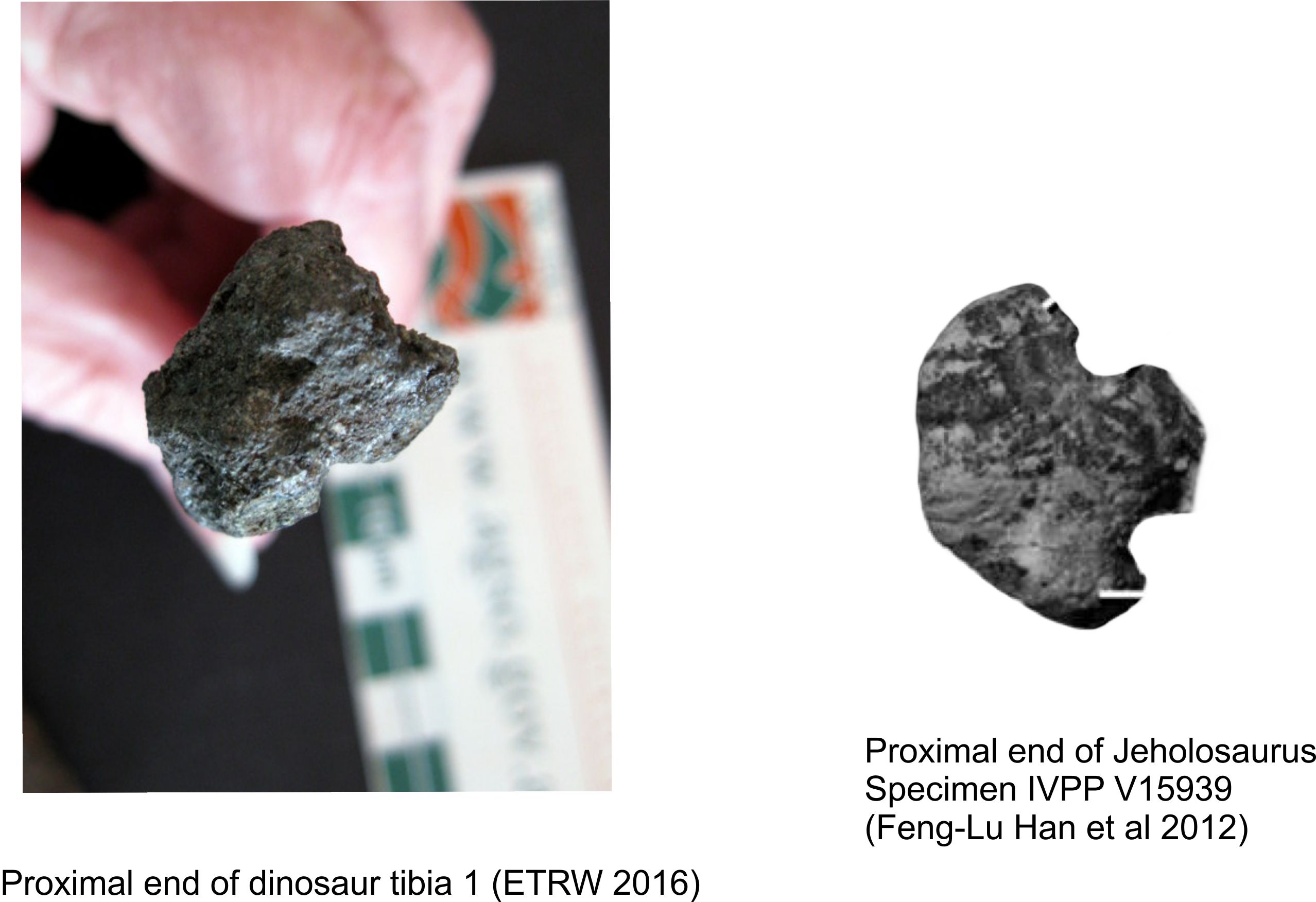
Image #7 tibia 1 from ETRW 2016.
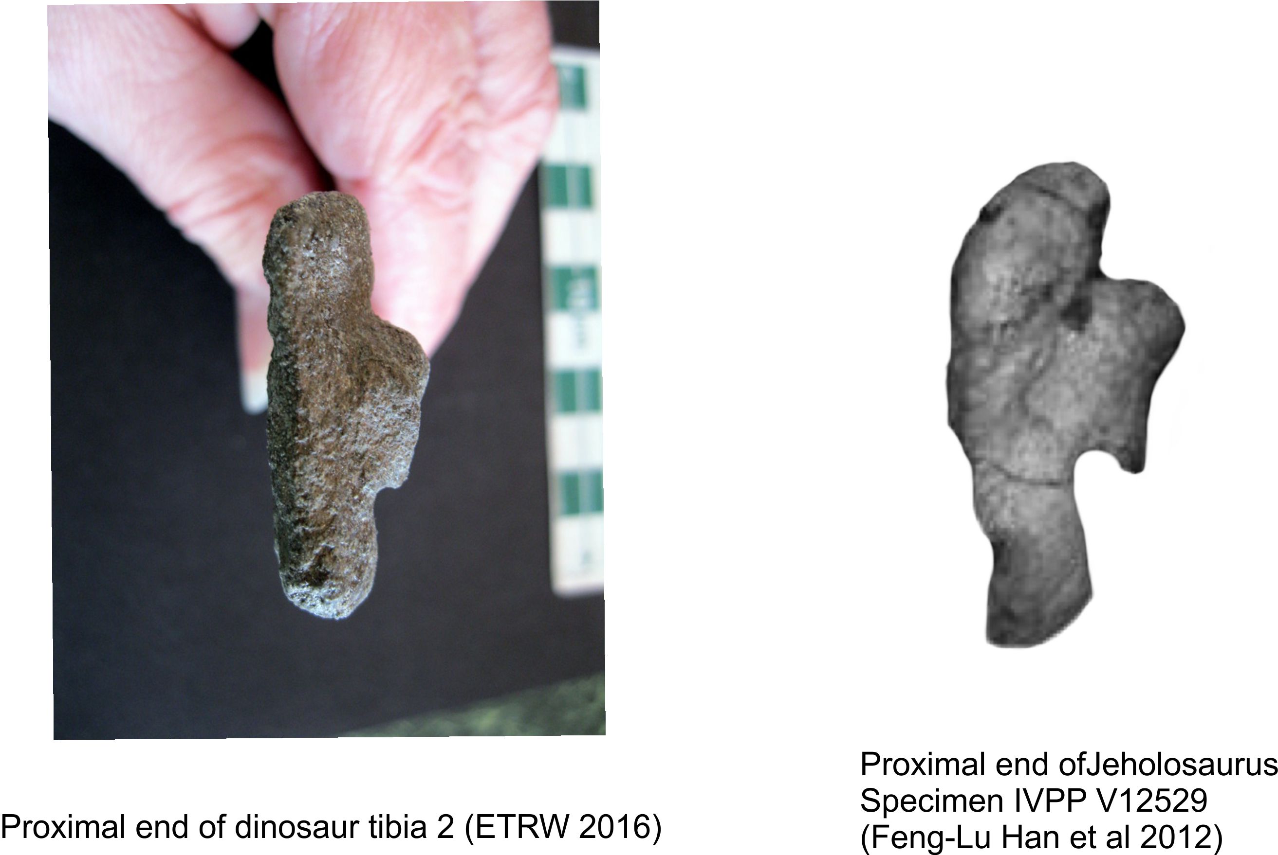
Image #8 tibia 2 from ETRW 2016.
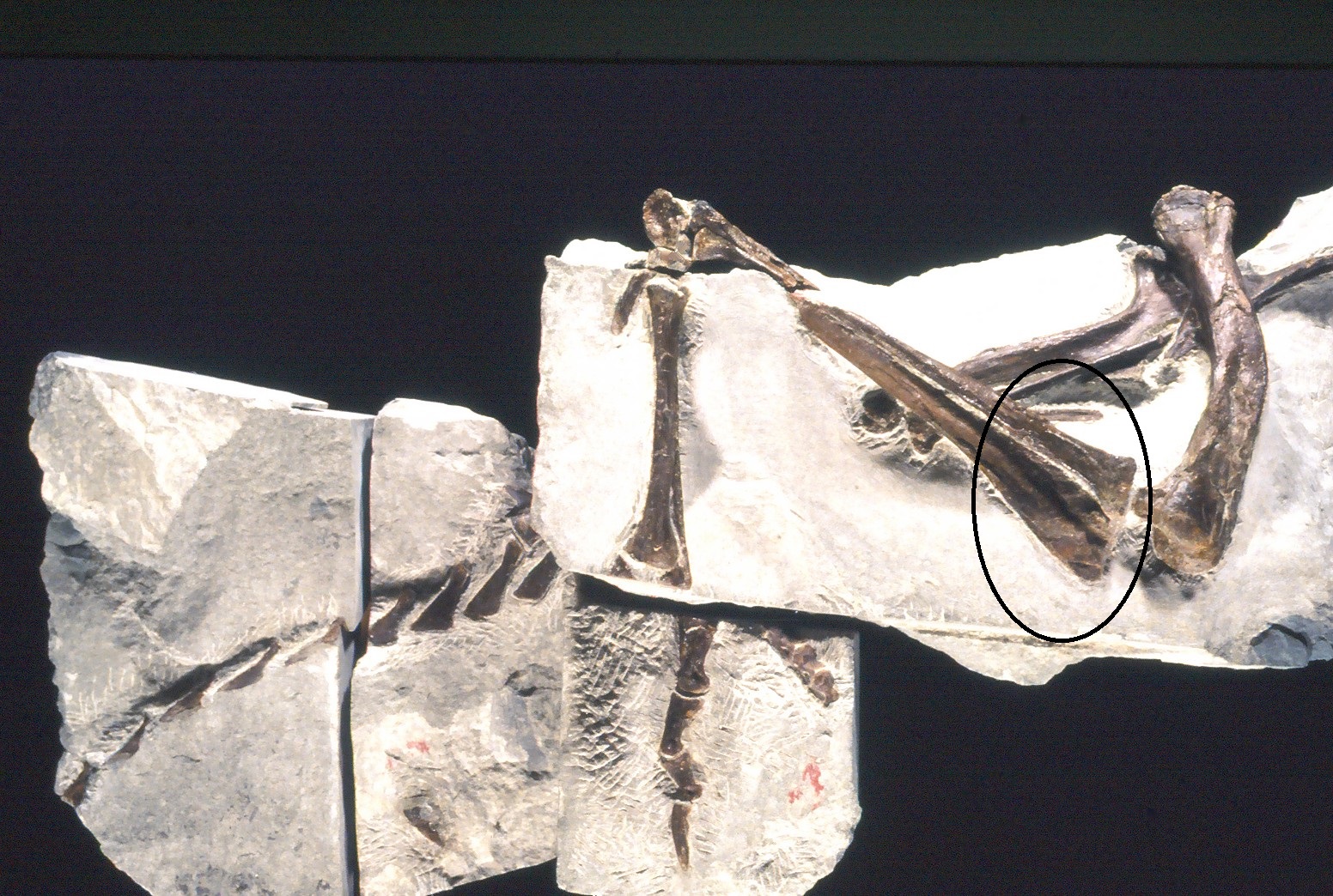
Image #9 - Partial skeleton from Dinosaur Cove.
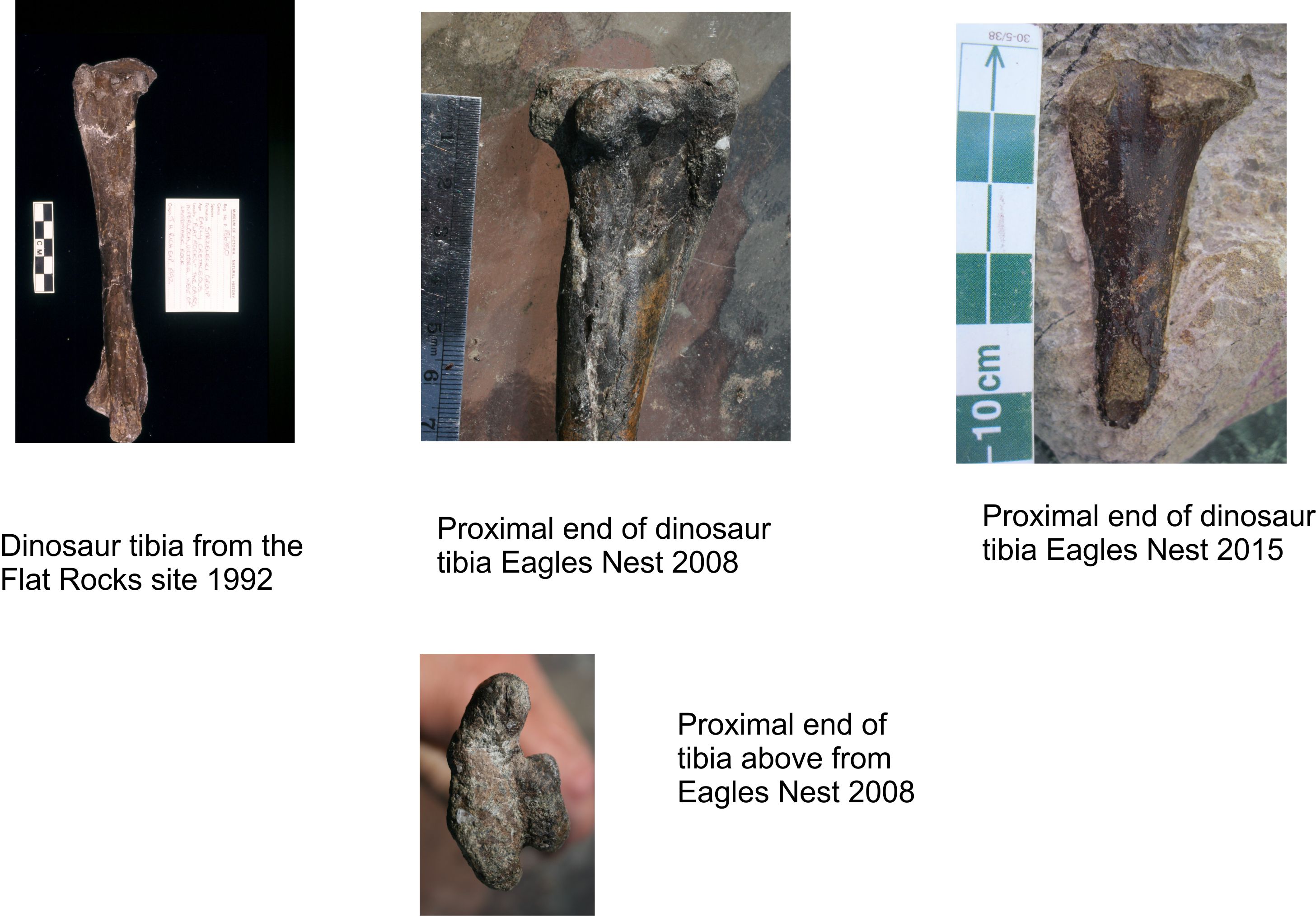
Image #10 tibiae from Bass Coast.
visits